Nail Matrix Function and Anatomy

The nail matrix is the area where your fingernails and toenails start to grow. Located at the base of the nail, it creates new cells that allow your nail to grow. Your nail may stop growing if the nail bed is injured.
The matrix creates new skin cells, which pushes out the old, dead skin cells to make your nails. As a result, injuries to the nail bed or disorders that affect the matrix can affect your nail growth.
Regarding the nail’s anatomy, it’s important to consider what you see and what you don’t. If you look at the top of the nail, you’re looking at the nail plate. Underneath the nail plate is the nail bed. The nail bed is where the nail adheres to the finger.
Other key elements of the nail include:
- Lunula. The white, half-moon cells at the nail’s base. Some people can only see the lunula on their thumbs while others cannot see theirs at all.
- Sterile matrix. This is the area of the nail above the lunula. The nail typically changes color beyond the germinal matrix (see below) as it extends to the sterile matrix because cells no longer have nuclei after that time, which makes the nail appear more transparent. This area is the next most common place where nail cells are made. Fingertip skin is connected to the sterile matrix.
- Germinal matrix. This is the area of the nail below the lunula (closest to the knuckle). An estimated 90 percent of nail production comes from the germinal matrix. This gives a natural curvature to the nail.
- Perionychium. The structures that surround the nail plate.
- Cuticle. The area of skin where the nail grows out of the finger. It provides protection to the nail matrix.
Your nails typically grow around 3 to 4 millimeters a month. Some people’s nails grow faster, including younger people and those with longer fingernails.
The nails are intended to provide protection to fingers as well as aid in opening, scratching, and tearing. Just like other body areas, they’re subject to injury and disease. The following are some conditions that can affect the nail matrix.
Trauma
An estimated 50 percentTrusted Source of fingernail injuries are due to a broken finger. Trauma to the nail can cause the production of new nail cells to stop for as long as three weeks.
Nail growth will usually resume at a faster rate and steady after about 100 days. You may notice the nail appears thicker than usual.
The extent of the injury often depends on where it occurs. If you have a deep cut or trauma to the germinal matrix at the base of the nail, it’s possible the nail may never grow back.
Ingrown nail
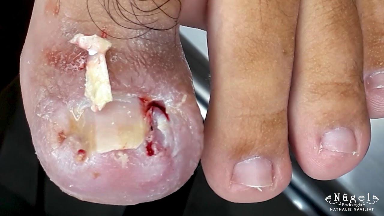
An ingrown nail occurs when a nail grows into the skin of the finger or toe, usually due to being cut too short. However, trauma to the nail and wearing tight shoes can also cause ingrown nails.
Symptoms include a swollen and tender nail. Sometimes, this area can get infected and will be red, painful, and sore.
Melanonychia
Melanonychia is a condition that causes brown pigmentation irregularities in the nail. Those who have dark skin are more likely to have it. This irregularity appears as a brown or black vertical stripe up the nail plate.
Melanonychia is a broad descriptive term that can indicate a normal variation on nail color or something as serious as subungual melanoma (see below). Several conditions and events can cause melanonychia, including:
Subungual melanoma
Subungual melanoma (or nail matrix melanoma) is a condition where cancerous cells grow in the nail matrix. The cancerous cells can cause changes in pigments in the nail known as melanin. As a result, a distinct striped discoloration can grow from the nail matrix.
If you observe changes to your nail that aren’t explained by trauma, talk to a doctor to ensure they’re not due to subungual melanoma.
Pterygium
Pterygium unguis is a condition that causes scarring that extends to the nail matrix. It causes the nail fold where the fingernail usually goes over the fingertip to fuse to the nail matrix. The nails take on a ridged appearance on the nail plate.
Lichen planus, burns, and lupus erythematosus cause pterygium.
Nevomelanocytic nevus
A nevomelanocytic nevus is essentially a mole or collection of melanocytes under the nail matrix. It’s possible to have one from birth, or acquire one following nail trauma or due to aging.
The challenge with a nevomelanocytic nevus is that it’s hard to tell the difference between a non-harmful nevus and discoloration that indicates cancer.
Paronychia
Paronychia is an infection of the fingernails or toenails. This condition may be acute or chronic, which can lead to nail deformities. Paronychia symptoms include swelling, redness, pain, and pus-filled areas in or around the nail. Fungus or bacteria can cause paronychia.
Dystrophic onychomycosis
Dystrophic onychomycosis is a fungal skin infection that causes total destruction of the nail plate. The condition usually occurs when a person has had a severe fungal nail infection for some time and goes untreated or isn’t fully treated.
Some common causes of dystrophic onychomycosis include:
- psoriasis
- lichen planus
- contact dermatitis
- trauma
A doctor can diagnose some nail concerns by a visual examination and listening to a description of symptoms. This is true for many fungal nail infections with nail crumbling, itching, and redness around the nail.
However, some conditions may warrant further work-up. This includes obtaining a specimen of the nail, either by clipping a portion of the end or performing a nail matrix biopsy.
Nail matrix biopsy
In a nail matrix biopsy, a doctor takes a sample of a nail matrix to examine for irregular cells, such as cancer. Because the nail matrix is deep at the nail’s base, doctors usually perform this procedure under local anesthesia.
A doctor can strategically inject local anesthetic into the finger’s base, numbing the finger. You shouldn’t be able to feel pain, only pressure, when a doctor removes a portion of the nail’s matrix. The approach to the biopsy depends on what area the doctor is testing.


