Favre-Racouchot Syndrome (Nodular Elastosis [Elastoidosis] with Cysts and Comedones, Senile or Solar Comedones, Smokers’ Comedones)

Are You Confident of the Diagnosis?
What you should be alert for in the history
Favre-Racouchot disease (FRD) is named after Favre and Racouchot, who described these lesions in 1932 and 1951, respectively.
Clinical history is significant for extensive ultraviolet light exposure.
Characteristic findings on physical examination
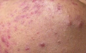
Clinical presentation consists of a slow and gradual development of 1-2mm non-inflammatory open and closed comedones, nodules, and cysts on a background of photodamaged and poikilodermatous skin (i.e. atrophic or thickened with a diffusely pale to yellowish hue, fine and/or deep rhytides and telangiectasia) on the face and neck (Figure 1).
Figure 1.
The periocular regions, malar eminence, temples, retroauricular and posterior and lateral neck (often in conjunction with cutis rhomboidalis nuchae) are most often affected. The lesions are bilateral unless there are specific circumstances where only one side of the face was sun-exposed (e.g. truck driver).
An uncommon related clinical and histologic variant of FRD is actinic comedonal plaque, which consists of a pink to bluish plaque with a cribriform appearance and clusters of deep open comedones, typically found on the flexural forearm. It is histologically indistinguishable from FRD. Other signs of actinic damage such as cutis rhomboidalis nuchae, actinic keratoses, and basal cell or squamous cell carcinoma can accompany FRD.
Expected results of diagnostic studies
Histopathologically, comedones of FRD appear as dilated pilosebaceous infundibulum filled with lamellar keratin and lined with flattened squamous epithelium with an extension to the surface. Often evident is a background of solar elastosis with epidermal atrophy and basophilic degeneration of the upper dermis, blue-gray discoloration of elastic fibers, as well as smaller, less numerous or absent sebaceous glands. Early FRD may show amorphous basophilic staining and elastic fiber hyperplasia, while later stages can be accompanied by dense collections of hypertrophic and entangled elastic fibers.
Diagnosis confirmation
The differential diagnosis for FRD includes a variety of conditions.
Actinic comedonal plaque affects flexural forearm. Actinic granuloma (synonym: annular elastolytic granuloma) features enlarging pink or skin-colored papules that form annular plaques with central atrophy in sun-exposed areas.
Acne vulgaris features open and closed comedones, papules, pustules, nodules, and cysts, typically in younger individuals without actinic skin changes.
Solar elastotic bands of the forearm presents with soft, yellow papules arranged in a cord-like distribution on flexural forearms, absence of open comedones, and is related to chronic sun exposure.
Cutis rhomboidalis nuchae features yellow, thickened skin with criss-crossed furrows on lateral and dorsal neck, and is related to chronic sun exposure
Milia can be distinguished by its yellowish to skin-colored papules, and it usually affects a younger age group.
Colloid milium presents with firm 1-5mm shiny smooth papules on the upper face, neck, ears, and dorsal hands, and is related to chronic sun exposure.
Syringoma features periocular skin-colored papules, and usually affects a younger age group.
Trichoepithelioma presents with skin-colored, dome-shaped papules, usually in the central face.
Chloracne is an acneiform eruption, consisting of comedones, pustules, papules, nodules, and cysts, most often in the postauricular region and cheeks, and includes a history of exposure to halogenated aromatic compounds.
Who is at Risk for Developing this Disease?
This condition is usually diagnosed in middle-aged Caucasian men (age 40-60) and affects up to 6% of all individuals over age 50. The primary risk factor is extensive ultraviolet light exposure from outdoor occupations (e.g. farmers, greenhouse workers, construction workers, plantation workers, truckers) and recreational activities (e.g. marathon runners, cyclists.) Smoking is also considered a risk factor.
What is the Cause of the Disease?
The exact pathophysiology of Favre-Racouchot disease (FRD) is unknown. It has been hypothesized that cumulative exposure to ultraviolet light (and cigarette smoking), among other external factors, results in progressive cutaneous atrophy and the formation of abnormal dermal elastic fibers (either by degeneration of existing elastic fibers or de novo formation of hypertrophic elastic fibers from overactive fibroblasts). Furthermore, disrupted keratinization of the pilosebaceous follicle and a dysfunctional elastic fiber support network may result in subsequent sebum retention and formation of comedones.
Ultraviolet light exposure may also produce increased free fatty acids and squalene, as well as squalene peroxidases, which may have comedogenic effects. In particular, ultraviolet-B light may promote sebaceous hyperplasia and increased sebum formation.
There may also be a genetic predisposition to this condition. Several cases of FRD following radiation therapy have been reported.
Medications have not been reported to exacerbate or cause FRD.
Systemic Implications and Complications
None
Treatment Options
Treatment options are summarized in Table I.
Table I.
| Medical Management | Surgical Management | Other Modalities |
|---|---|---|
| Topical retinoids and related derivatives for daily use: tretinoin, retinoic acid, retinaldehyde (less irritation), tazarotene (most irritation), adapalene (synthetic retinoid, less irritation) | Manual extraction with comedone extractor | Dermabrasion |
| Chemical peels–start with 50-70% glycolic acid (superficial peel) and if tolerated or if no improvement is noted, progress to a 15% trichloroacetic acid (medium-depth peel), and very cautiously proceed to phenol (deep peel) if indicated. | Curettage | Carbon dioxide laser |
| Systemic: low-dose isotretinoin 0.05-1mg/kg/day | Note: The combination of surgical and medical therapy yields maximal benefit | Fractionated laser |
| Alpha-hydroxy acids | ||
| Lifestyle modification–sun protection, smoking cessation, although this will not reverse preexisting actinic damage | ||
| Periodic skin assessment for skin cancer |
Optimal Therapeutic Approach for this Disease
The treatment of choice is manual extraction, followed by topical retinoids or their derivatives. Large comedones require manual extraction and are unlikely to respond to topical treatment alone.
Retinoids work by normalizing keratinization, promoting exfoliation and comedone extraction, and improving dermal elastic fiber and collagen synthesis. Examples of such products include topical tretinoin, retinoic acid, and retinaldehyde (precursor to retinoic acid, considered less irritating).
Topical therapies require several months of continuous use before improvement is noted. A suggested regimen would be tretinoin 0.04% gel daily, and if tolerated, the strength may be increased to 0.1%. Retinoids in a cream-based vehicle or retinaldehyde are recommended for more sensitive skin.
Other alternative therapies include manual extraction or surgical excision alone (for a small number of lesions), chemical peels, dermabrasion, and resurfacing lasers, with or without topical retinoids. A carbon dioxide laser has been used safely and effectively using two initial passes, followed by manual extraction of comedones, and then a third pass in fifty patients with Fitzpatrick skin type III, with no evidence of relapse 15 to 21 months later (see Mavilia et al. 2010). Oral isotretinoin given as 0.05 to 1mg/kg/day over 4 to 5 months, with or without topical retinoids may also be used.
Patient Management
Daily treatment with topical retinoids, as tolerated. Use should be decreased if irritation from the medication occurs. These patients often have extensive actinic damange and it would be important to monitor them periodically for skin cancer.
Unusual Clinical Scenarios to Consider in Patient Management
Unusual clinical scenarios regarding FRD include the association of cutaneous myxomas without evidence of the Carney complex; on rare occasions, mycosis fungoides may affect follicular and eccrine structures to mimic FRD.
What is the Evidence?
Aloi, F, Tomasini, C, Pippione, M. “mycosisfungoides and eruptive epidermoid cysts: a unique response of follicular and eccrine structures”. Dermatology. vol. 187. 1993. pp. 273-7. (A case report of one patient with mycosisfungoides, presenting with small eruptive cutaneous cysts on the face, neck, and upper trunk, reminiscent of FRD. Mycoses fungoides can appear as cyst-like lesions via alterations in the development of follicular and eccrine structures. Histology was required to make the diagnosis of mycosis fungoides.)
Benlier, E, Alicioglu, B, Kandulu, H, Yurdakul, ES, Top, H. “An unusual association of cutaneous myxoma with Favre-Racouchot syndrome”. Prague Med Rep. vol. 109. 2008. pp. 321-4. (The first case report of multiple cutaneous myxomas on the feet associated with Favre-Racouchot syndrome.)
Favre, M. “Sur une affection kystique des appareils pilo-sébacés localisée à certaines régions de la face”. Bull Soc Fr Dermatol Syph. vol. 39. 1932. pp. 93-6.
Favre, M, Racouchot, J. “L’elasteidose cutanée nodulaire a kystes et a comedons”. Ann Dermatol Syph. vol. 78. 1951. pp. 681-702.
Breit, S, Flaig, MJ, Wolff, H, Plewig, G. “Favre-Racouchot-like disease after radiation therapy”. J Am Acad Dermatol. vol. 49. 2003. pp. 117-9. (A case report of a 62-year-old female who developed Favre-Racouchot-like comedones two months postradiation therapy to the nasopharynx. Treatment with low-dose isotretinoin 20mg/day, isotretinoin gel, and multiple courses of manual extraction over a five-month period resulted in significant improvement in cosmesis without recurrence.)
Friedman, SJ, Su, WP. “Favre-Racouchot syndrome associated with radiation therapy”. Cutis. vol. 31. 1982. pp. 306-10. (A case report of a 56-year-old female who developed Favre-Racouchot syndrome on her face and scalp three months postradiation therapy of an astrocytoma. Treatment with topical retinoic acid gel yielded an excellent outcome.)
Keough, GC, Laws, RA, Elston, DM. “Favre-Racouchot syndrome: a case of smokers’ comedones”. Arch Dermatol. vol. 133. 1997. pp. 796-7. (A case series of 57 patients with Favre-Racouchot disease [mean age 66.4 years], compared to 307 consecutive controls [mean age 67.8 years] without the condition, from a cancer screening clinic in Texas. The majority of the patients were Caucasian retired military personnel. Smokers were defined as those with greater than 5 pack years. Favre-Racouchot disease was more often found in smokers [21%] compared to non-smokers [9.9%].)
Motoyoshi, K. “Enhanced comedo formation in rabbit ear skin by squalene and oleic acid peroxidases”. Br J Dermatol. vol. 98. 1983. pp. 109-91. (Various potentially comedogenic substances such as squalene, squalene peroxidases, oleic acid, and oleic acid peroxidases were tested on the rabbit ear model. Ultraviolet light A was used to produce peroxidase products. Levels of these comedogenic substances were measured and the skin was sampled for histology evaluation. Epithelial hyperplasia and hyperkeratosis within the follicular infundibulum and sebaceous gland proliferation were documented. Squalene peroxidases (not squalene), oleic acid, and oleic acid peroxidases were associated with large comedones and were considered comedogenic.)
Sánchez-Yus, E, del Rio, E, Simón, P, Requena, L, Vázquez, H. “The histopathology of closed and open comedones of Favre-Racouchot disease”. Arch Dermatol. vol. 133. 1997. pp. 742-5. (A case series involving eight patients [nineteen cysts and comedones]. Comedones of Favre-Racouchot disease demonstrated marked solar elastosis in the dermis, but otherwise had identical features to acne vulgaris, including the presence of abundant bacteria. Unlike acne vulgaris, all cysts and comedones except for one contained vellus type hairs.)
Mavilia, L, Campolmi, P, Santoro, G, Lotti, T. “Combined treatment of Favre-Racouchot syndrome with a superpulsed carbon dioxide laser: report of 50 cases”. Dermatol Ther. vol. 23. 2010. pp. S4-S6. (A case series of fifty patients [age 55-71, mean age 60 years] with Fitzpatrick III skin type, treated with superpulsed carbon dioxide laser [Smartoffice, DEKA M.E.L.A., Florence, Italy] with parameters 1-2% duty-cycle, 10Hz frequency and 2-3mm spot size. Carbon dioxide laser was used for epidermal vaporization [two passes] and followed by manual extraction of comedones with forceps and another pass of carbon dioxide laser. Safety and efficacy with minimal adverse effects and rapid recovery were demonstrated in all patients.)
Patterson, WM, Fox, MD, Schwartz, RA. “Favre-Racouchot disease”. Int J Dermatol. vol. 43. 2004. pp. 167-9. (A brief, comprehensive review article on the epidemiology, pathogenesis, clinical presentation, and management of Favre-Racouchot disease.)
Rallis, E, Karanikola, E, Verros, C. “Successful treatment of Favre-Racouchot disease with 0.05% tazarotene gel”. Arch Dermatol. vol. 143. 2007. pp. 810-2. (A case series of three patients with Favre-Racouchot disease, treated with tazarotene 0.05% gel daily for a mean 7.5 weeks. Improvement was noted in all patients. Therefore, topical tazarotene may be considered, particularly if other topical retinoids have been ineffective and there are barriers for surgical or resurfacing treatment modalities.)
Sharkey, MJ, Keller, RA, Grabski, WJ, McCollough, ML. “Favre-Racouchot syndrome. A combined therapeutic approach”. Arch Dermatol. vol. 128. 1992. pp. 615-6. (A case report of a 57-year-old woman with massive nodular cystic lesions on bilateral cheeks, which were histologically in keeping with Favre-Racouchot sydrome. The authors performed a multiple-staged excision to debulk the malar masses followed by dermabrasion and daily tretinoin cream. No recurrence was reported
Copyright © 2017, 2013 Decision Support in Medicine, LLC. All rights reserved.
No sponsor or advertiser has participated in, approved or paid for the content provided by Decision Support in Medicine LLC. The Licensed Content is the property of and copyrighted by DSM.


