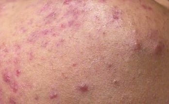
persistent cutaneous abscesses
- Nosocomial cutaneous abscesses in septic infants.
Mandel, D; Littner, Y; Mimouni, F B; Dollberg, S
To retrospectively study the epidemiology of nosocomial cutaneous abscesses in 46 consecutive septic infants. Ten infants had one abscess or more. Surviving infants with abscesses had a longer duration of bacteraemia, which disappeared within 24 hours of drainage. Infants with persistent bacteraemia should be examined regularly for the presence of abscesses.
- [Aseptic cutaneous breast abscesses associated with ulcerative colitis].
Sallé de Chou, C; Ortonne, N; Hivelin, M; Wolkenstein, P; Chosidow, O; Valeyrie-Allanore, L
2016-02-01
Inflammatory bowel diseases are associated with a broad range of cutaneous lesions. Herein we report the first case of aseptic skin abscesses associated with ulcerative colitis. Since March 2008, a 40-year-old woman presented with bilateral mammary abscesses, relapsing despite repeated antibiotic treatment. She was followed for ulcerative colitis diagnosed in 2011 by means of a rectal biopsy. Despite four surgical procedures, there was no improvement in her mammary abscesses and bilateral mastectomy was then proposed because of the persistent symptoms. Her general state of health remained stable. Clinically, there were bilateral inflammatory nodes with fistulae and pus. These lesions were extremely painful. Mild inflammatory syndrome was noted, but the immunological tests revealed nothing of note. Bacteriological, parasitological and mycological tests on biopsy specimens were negative. Histological examination of a surgical biopsy revealed lymphoplasmacytic infiltration of the dermis and subcutis with altered polymorphonuclear cells and epithelioid granuloma. The CT-scan showed no other remote lesions. The final diagnosis was cutaneous aseptic abscess syndrome associated with ulcerative colitis. Colchicine 1mg/day was initiated and resulted in regression of the skin lesions, with complete remission at one year of follow-up. Aseptic abscess syndrome must be considered in the event of recurrent aseptic cutaneous abscesses which may be associated with inflammatory bowel disease. Surgery should be avoided and treatment should be based on suitable drug therapy. Copyright © 2016. Published by Elsevier Masson SAS.
- Skin abscess
Abscess – skin; Cutaneous abscess; Subcutaneous abscess; MRSA – abscess; Staph infection – abscess … Skin abscesses are common and affect people of all ages. They occur when an infection causes pus …
- Primary versus secondary closure of cutaneous abscesses in the emergency department: a randomized controlled trial.
Singer, Adam J; Taira, Breena R; Chale, Stuart; Bhat, Rahul; Kennedy, David; Schmitz, Gillian
2013-01-01
Cutaneous abscesses have traditionally been treated with incision and drainage (I&D) and left to heal by secondary closure. The objective was to compare the healing rates of cutaneous abscesses following I&D after primary or secondary closure. This was a randomized, controlled, trial, balanced by center, with blocked randomization created by a random-number generator. One urban and one suburban academic emergency department (ED) participated. Subjects were randomized to primary or secondary wound closure following I&D of the abscess. Main outcome measures were the percentage of healed wounds (wound was completely closed by visual inspection; a 40% difference in wound healing was sought) and overall failure rate (need for additional intervention including suture removal, additional drainage, antibiotics, or admission within 7 days after drainage). Fifty-six adult patients with simple localized cutaneous abscesses were included; 29 were randomized to primary closure, and 27 were randomized to secondary closure. Healing rates at 7 days were similar between the primary and secondary closure groups (69.6%, 95% confidence interval [CI] = 49.1% to 84.4% vs. 59.3%, 95% CI = 40.7% to 75.5%; difference 10.3%, 95% CI = -15.8% to 34.1%). Overall failure rates at 7 days were also similar between the primary and secondary closure groups (30.4%, 95% CI = 15.6% to 50.9% vs. 28.6%, 95% CI = 15.2% to 47.1%; difference 1.8%, 95% CI = -24.2% to 28.8%). The rates of wound healing and treatment failure following I&D of simple abscesses in the ED are similar after primary or secondary closure. The authors did not detect a difference of at least 40% in healing rates between primary and secondary closure. © 2013 by the Society for Academic Emergency Medicine.
- Cutaneous human papillomaviruses persist on healthy skin.
Hazard, Kristina; Karlsson, Anna; Andersson, Kristin; Ekberg, Henrik; Dillner, Joakim; Forslund, Ola
2007-01-01
Cutaneous human papillomaviruses (HPVs) are frequently found in healthy skin and have also been implicated in non-melanoma skin cancer. For genital HPV types, a persistent infection with one of the high-risk types is a prerequisite for the development of cervical cancer. However, there is only limited data on whether infections with cutaneous HPV types persist over time. Serial forehead swab samples collected from 63 volunteers (42 healthy individuals and 31 renal transplant recipients (RTRs)), sampled 6.3 years (range: 5.0-7.0 years) apart, were analyzed for HPV using general primer PCR, cloning, and sequencing. Among the healthy individuals, the prevalences of HPV were 69% (29/42) at enrolment and 71% (30/42) at follow-up. Among the individuals positive at baseline, 48% (14/29) had a persistent infection. Among the RTRs, 71% (15/21) were positive for HPV at enrolment and 90% (19/21) at follow-up. A persistent infection was detected in 33% (5/15). In total, HPV was detected in 44 of the samples collected at baseline and the same virus was found at follow-up in 43% (19/44). Persistence was not significantly associated with age, sex, immunosuppressive treatment, history of warts, or genus of HPV. We conclude that cutaneous HPV infections commonly persist over several years on healthy skin.
- Identification and molecular characterization of Corynebacterium xerosis isolated from a sheep cutaneous abscess: first case report in Mexico.
Hernández-León, Fernando; Acosta-Dibarrat, Jorge; Vázquez-Chagoyán, Juan Carlos; Rosas, Pomposo Fernandez; de Oca-Jiménez, Roberto Montes
2016-07-22
Corynebacterium xerosis is a commensal organism found in skin and mucous membranes of humans. It is considered an unusual pathogen, and it is rarely found in human and animal clinical samples. Here we describe the isolation of C. xerosis from a 4-months-old Pelifolk lamb located in Tesistán, central western Mexico. This microorganism should be considered for differential diagnosis in cutaneous abscessed lesions in sheep, as it represents a zoonotic risk factor for human infection in sheep farms. The animal exhibited a hard-consistency, 5 cm diameter abscess, without drainage, in the neck. The presumptive clinical diagnosis was caseous lymphadenitis, caused by Corynebacterium pseudotuberculosis. Samples were obtained by puncture and cultured in 8 % sheep blood agar under microaerophilic conditions. Colonies were non-haemolytic, brown-yellowish and showed microscopic and biochemical features similar to C. pseudotuberculosis, except for the urea test. A multiplex-PCR for the amplification of partial sequences of the pld, rpoB and intergenic fragment from 16S to 23S genes suggested that isolate could be C. xerosis, which was later confirmed by sequencing analysis of the rpoB gene. This study shows for the first time isolation and molecular characterization of C. xerosis from a clinical sample of an ovine cutaneous abscess in Mexico. This finding highlights the need for differential diagnosis of this pathogen in ovine skin abscesses, as well as epidemiological and control studies of this pathogen in sheep farms.
- [Topical treatment of persistent cutaneous leishmaniasis with paromomycin].
Flaig, M J; Rupec, J; Ruzicka, T; Rupec, R A
2007-08-01
Cutaneous leishmaniasis is an infectious disease with increasing prevalence in Germany. Diagnosis and therapy may be difficult due to the variability of the clinical and histomorphological picture and resistance to therapy. In this case study we report on a female patient with a persistent cutaneous leishmaniasis successfully treated with topical administration of paromomycin.
- Intracardiac abscess with cutaneous fistula secondary to ventricular septal defect repair simulating sternal wound infection.
Rafael, Aldo Elmer; Keshavamurthy, Suresh; Sepulveda, Edgardo; Miranda, Cyndee Cruz; Okamoto, Toshihiro; Pettersson, Gosta Bengt
2014-06-01
Cutaneous fistula as a clinical presentation of intracardiac abscess of the right side is such an unusual occurrence that it has not until now been reported in the English-language medical literature. We present a rare case of right-sided infective endocarditis caused by Achromobacter xylosoxidans in which recurrent infection presented as sternal wound discharge. The infection was found to have an intracardiac origin and was successfully managed by radical débridement on cardiopulmonary bypass.
- Intracardiac Abscess with Cutaneous Fistula Secondary to Ventricular Septal Defect Repair Simulating Sternal Wound Infection
Keshavamurthy, Suresh; Sepulveda, Edgardo; Miranda, Cyndee Cruz; Okamoto, Toshihiro; Pettersson, Gosta Bengt
2014-01-01
Cutaneous fistula as a clinical presentation of intracardiac abscess of the right side is such an unusual occurrence that it has not until now been reported in the English-language medical literature. We present a rare case of right-sided infective endocarditis caused by Achromobacter xylosoxidans in which recurrent infection presented as sternal wound discharge. The infection was found to have an intracardiac origin and was successfully managed by radical débridement on cardiopulmonary bypass. PMID:24955054
- Ultrasound Visualization of Atypical Abscess Ultimately Containing Bot Fly Larva.
Bovino, Patrick; Cole, John; Scheatzle, Mark
2016-08-01
Because of the rise in community-acquired methicillin-resistant Staphylococcus aureus (CA-MRSA), presentations to the emergency department for the evaluation of cutaneous abscesses have risen dramatically over the past 2 decades. Soft tissue point of care ultrasound (POCUS) differentiates abscess from cellulitis, determines the size and shape, and characterizes the contents of the abscess. It has been shown to improve medical decision-making and therefore the emergency management of cutaneous abscesses over physical examination alone. We report a case of an unusual nonhealing abscess in an 18-year-old woman with a recent history of foreign travel where soft tissue POCUS identified motion within the abscess pocket. This changed the management of the case, leading to the diagnosis of bot fly myiasis. WHY SHOULD AN EMERGENCY PHYSICIAN BE AWARE OF THIS?: Clinicians should entertain a broader differential for an apparent abscess and consider liberal use of soft tissue POCUS in these cases. Copyright © 2016 Elsevier Inc. All rights reserved.
- Abscess incision and drainage in the emergency department–Part I.
Halvorson, G D; Halvorson, J E; Iserson, K V
1985-01-01
Superficial abscesses are commonly seen in the emergency department. In most cases, they can be adequately treated by the emergency physician without hospital admission. Treatment consists of surgical drainage with the addition of antibiotics in selected cases. Incision is generally performed using local anesthesia, with intraoperative and postoperative systemic analgesia. Care must be taken to make a surgically appropriate incision that allows adequate drainage without injuring important structures. Postoperative care includes warm soaks, drains or wicks, analgesia, and close follow-up. Antibiotics are usually unnecessary. Complications of incision and drainage include damage to adjacent structures, bacteremic complications, misdiagnosis of such entities as mycotic aneurysms, and spread of infection owing to inadequate drainage. The infectious agents responsible for abscess formation are numerous and depend largely on the anatomic location of the abscess. Staphylococcus aureus accounts for less than half of all cutaneous abscesses. Anaerobic bacteria are common etiologic agents in the perineum and account for the majority of all cutaneous abscesses. Abscesses at specific locations involve special consideration for diagnosis and treatment and may require specialty consultation.
- Persistent parasites and immunologic memory in cutaneous leishmaniasis: implications for vaccine designs and vaccination strategies.
Okwor, Ifeoma; Uzonna, Jude
2008-01-01
Despite a plethora of publications on the murine model of cutaneous leishmaniasis and their contribution to our understanding of the factors that regulate the development of CD4+ T cell immunity in vivo, there is still no effective vaccine against the human disease. While recovery from natural or experimental infection with Leishmania major, the causative agent of human cutaneous leishmaniasis, results in persistence of parasites at the primary infection site and the development of long-lasting immunity to reinfection, vaccination with killed parasites or recombinant proteins induces only short-term protection. The reasons for the difference in protective immunity following recovery from live infection and vaccination with heat-killed parasites are not known. This may in part be related to persistence of live parasites following healing of primary cutaneous lesions, because complete clearance of parasites leads to rapid loss of infection-induced immunity. Recent reports indicate that in addition to persistent parasites, IL-10-producing natural regulatory T cells may also play critical roles in the maintenance and loss of infection-induced immunity. This review focuses on current understanding of the factors that regulate the development, maintenance and loss of anti-Leishmania memory responses and highlights the role of persistent parasites and regulatory T cells in this process. Understanding these factors is crucial for designing effective vaccines and vaccination strategies against cutaneous leishmaniasis.
- Ampicillin/sulbactam and cefoxitin in the treatment of cutaneous and other soft-tissue abscesses in patients with or without histories of injection drug abuse.
Talan, D A; Summanen, P H; Finegold, S M
2000-08-01
A randomized, double-blind trial compared the clinical and bacteriologic efficacy of ampicillin/sulbactam (2 g/1 g) and cefoxitin (2 g) administered intravenously every 6 h to patients with (n=49) or without (n=47) histories of injection drug abuse who presented with cutaneous or other soft-tissue infections. Cure or improvement occurred in 89.8% of ampicillin/sulbactam-treated patients, compared with 93.6% of cefoxitin-treated patients. The median time to resolution of all symptoms was 10.5 days with ampicillin/sulbactam treatment and 15.5 days with cefoxitin treatment. Mixed aerobic-anaerobic infection was encountered frequently in both treatment groups. A significantly higher percentage of Streptococcus species was found in the major abscesses of the patients with histories of injection drug abuse, compared with those without such histories (37% vs. 19%, respectively; P=.0009). Overall, ampicillin/sulbactam eradicated pathogens from the major abscesses in 100% of patients, whereas the eradication rate with cefoxitin was 97.9%. The 2 drugs were well tolerated. Ampicillin/sulbactam and cefoxitin were equally effective for the empirical treatment of cutaneous or other soft-tissue infections in injection drug abusers and patients who did not inject drugs.
- Understanding the Lung Abscess Microbiome: Outcomes of Percutaneous Lung Parenchymal Abscess Drainage with Microbiologic Correlation.
Duncan, Christopher; Nadolski, Gregory J; Gade, Terence; Hunt, Stephen
2017-06-01
Lung parenchymal abscesses represent an uncommon pathology with high mortality if untreated. Although most respond well to antibiotics, the optimal therapy for persistent abscesses is unknown. The purpose of this study was to review the outcomes of percutaneous lung parenchymal abscess catheter drainage after broad-spectrum antibiotic therapy failure and correlate with patient microbiologic samples. Retrospective review of patients who underwent percutaneous lung abscess drainage at a tertiary hospital system from 2005 to 2015 was performed. In total, 19 procedures were identified on 16 different patients; six females and ten males. Mean patient age was 55 years (range 22-81). Median follow-up time was 7 months (range <1-78). Technical success was 100%. There was one major complication, a pneumothorax. Follow-up was until tube removal or death in 100% of patients. Catheters were removed with resolution of the abscess cavity in 58% (11/19) or with non-draining abscess cavities in 21% (4/19) for a clinical success rate of 79%. Blood cultures demonstrated no growth in all cases, while 21% (4/19) of sputum or bronchoscopic cultures demonstrated growth. In comparison, the specimens from initial catheter placement isolated a causative organism in 95% (18/19) of case (p < 0.0001). In cases of persistent lung abscess after broad-spectrum antibiotics, percutaneous abscess drainage is highly sensitive for microbiologic sampling compared to sputum/bronchoscopic or blood cultures. Additionally, percutaneous drainage of lung parenchymal abscess cavities may promote resolution of the abscess with high rates of therapeutic success and low complications.
- Cutaneous community-acquired methicillin-resistant Staphylococcus aureus infection in participants of athletic activities.
Cohen, Philip R
2005-06-01
Cutaneous community-acquired methicillin-resistant Staphylococcus aureus (CAMRSA) has been identified in otherwise healthy individuals either with or without methicillin-resistant S. aureus (MRSA)-associated risk factors who participate in athletic activities. The purpose of this study was to describe the clinical features of CAMRSA skin infection that occurred in university student athletes, evaluate the potential mechanisms for the transmission of MRSA infection of the skin in participants of athletic activities, and review the measures for preventing the spread of cutaneous CAMRSA infection in athletes. A retrospective chart review of the student athletes from the University of Houston whose skin lesions were evaluated at the Health Center and grew MRSA was performed. The clinical characteristics and the postulated mechanisms of cutaneous MRSA infection in the athletes were compared with those previously published in reports of CAMRSA skin infection outbreaks in other sports participants. Cutaneous CAMRSA infection occurred in seven student athletes (four women and three men) who were either weight lifters (three students) or members of a varsity sports team: volleyball (two women), basketball (one woman), and football (one man). The MRSA skin infection presented as solitary or multiple, tender, erythematous, fluctuant abscesses with surrounding cellulitis. The lesions were most frequently located in the axillary region (three weight lifters), on the buttocks (two women), or on the thighs (two women). The drainage from all of the skin lesions grew MRSA, which was susceptible to clindamycin, gentamicin, rifampin, trimethoprim/sulfamethoxazole, and vancomycin; five of the isolates were also susceptible to ciprofloxacin and levofloxacin. All of the bacterial strains were resistant to erythromycin, oxacillin, and penicillin. The cutaneous MRSA infections persisted or worsened in the six athletes who were empirically treated for methicillin-sensitive S. aureus at
- Understanding the Lung Abscess Microbiome: Outcomes of Percutaneous Lung Parenchymal Abscess Drainage with Microbiologic Correlation
DOE Office of Scientific and Technical Information (OSTI.GOV)
Duncan, Christopher; Nadolski, Gregory J.; Gade, Terence
IntroductionLung parenchymal abscesses represent an uncommon pathology with high mortality if untreated. Although most respond well to antibiotics, the optimal therapy for persistent abscesses is unknown. The purpose of this study was to review the outcomes of percutaneous lung parenchymal abscess catheter drainage after broad-spectrum antibiotic therapy failure and correlate with patient microbiologic samples.Materials and MethodsRetrospective review of patients who underwent percutaneous lung abscess drainage at a tertiary hospital system from 2005 to 2015 was performed. In total, 19 procedures were identified on 16 different patients; six females and ten males. Mean patient age was 55 years (range 22–81). Median follow-upmore » time was 7 months (range <1–78).ResultsTechnical success was 100%. There was one major complication, a pneumothorax. Follow-up was until tube removal or death in 100% of patients. Catheters were removed with resolution of the abscess cavity in 58% (11/19) or with non-draining abscess cavities in 21% (4/19) for a clinical success rate of 79%. Blood cultures demonstrated no growth in all cases, while 21% (4/19) of sputum or bronchoscopic cultures demonstrated growth. In comparison, the specimens from initial catheter placement isolated a causative organism in 95% (18/19) of case (p < 0.0001).ConclusionIn cases of persistent lung abscess after broad-spectrum antibiotics, percutaneous abscess drainage is highly sensitive for microbiologic sampling compared to sputum/bronchoscopic or blood cultures. Additionally, percutaneous drainage of lung parenchymal abscess cavities may promote resolution of the abscess with high rates of therapeutic success and low complications.« less
- Bone formation within a breast abscess
Mannu, Gurdeep Singh; Ahmed, Farid; Cunnick, Giles; Mungalsingh, Naren
2014-01-01
We present a rare case of osseous metaplasia in a poorly healing breast abscess. An 87-year-old woman was referred to the breast surgery clinic with a painful lump in her right breast. Initial imaging and core biopsy suggested a breast abscess. Despite several courses of antibiotics and repeated attempts at aspiration the painful lesion persisted. It was eventually surgically excised in its entirety and final histopathology showed the presence of bone formation within the abscess. The patient’s symptoms subsequently resolved. To the best of our knowledge, this is the first case in the literature, of osseous metaplasia within a breast abscess in the absence of malignancy. PMID:25246453
- Bone formation within a breast abscess.
Mannu, Gurdeep Singh; Ahmed, Farid; Cunnick, Giles; Mungalsingh, Naren
2014-09-22
We present a rare case of osseous metaplasia in a poorly healing breast abscess. An 87-year-old woman was referred to the breast surgery clinic with a painful lump in her right breast. Initial imaging and core biopsy suggested a breast abscess. Despite several courses of antibiotics and repeated attempts at aspiration the painful lesion persisted. It was eventually surgically excised in its entirety and final histopathology showed the presence of bone formation within the abscess. The patient’s symptoms subsequently resolved. To the best of our knowledge, this is the first case in the literature, of osseous metaplasia within a breast abscess in the absence of malignancy. 2014 BMJ Publishing Group Ltd.
- [A case of cutaneous protothecosis].
Kazantseva, I A; Molochkov, A V; Sukhov, A V; Bondarenko, E V
The paper describes a case of a rare opportunistic infection, such as skin lesion caused by achlorophyllic unicellular algae of the genus Prototheca. It provides a detailed pathologic description of the foci of cutaneous protothecosis, such as pandermal inflammatory infiltrate, granulomas, pseudoepitheliomatous hyperplasia, and intraepidermal abscesses. Criteria for pathogen detection in histological sections are given.
- Clinical patterns of cutaneous nontuberculous mycobacterial infections.
Bartralot, R; GarcÃa-Patos, V; Sitjas, D; RodrÃguez-Cano, L; Mollet, J; MartÃn-Casabona, N; Coll, P; Castells, A; Pujol, R M
2005-04-01
Cutaneous nontuberculous mycobacterial infections result from external inoculation, spread of a deeper infection, or haematogenous spread of a disseminated infection. There are two species-specific infections (fish-tank or swimming-pool granuloma, due to Mycobacterium marinum, and Buruli ulcer, caused by M. ulcerans). Most infections, however, produce a nonspecific clinical picture. To define clinical patterns of cutaneous disease in nontuberculous mycobacterial infections. Fifty-one patients with cutaneous nontuberculous mycobacterial infections were reviewed. Clinical and histopathological features of normal hosts and immunosuppressed patients were compared. Two subgroups of immunosuppressed patients were distinguished: patients with cutaneous infection and patients with a disseminated infection and cutaneous involvement. In immunosuppressed patients the number of lesions was significantly higher. Abscesses and ulceration were also more frequently observed. Different species were found in normal hosts and immunosuppressed patients. Several clinical patterns of cutaneous infection were defined: lymphocutaneous or sporotrichoid lesions; nonlymphocutaneous lesions at the site of trauma; folliculitis and furunculosis involving the lower extremities; disseminated lesions on the extremities in immunosuppressed patients. Two patterns were observed in patients with a disseminated infection: localized cutaneous lesions and disseminated cutaneous and mucosal lesions. Cutaneous manifestations of nontuberculous mycobacterial infections may be classified according to criteria such as cutaneous lesions and immune status.